

Blog / Pain Science
What Your MRI Doesn't Tell You About Your Back Pain
Bulging discs, degeneration and 'wear and tear' show up on scans of people with no pain at all. Here is what that means for your recovery.
August 2026 · 7 min read
You have had back pain for a few months. You finally get a scan. The report comes back with words like disc degeneration, disc bulge, annular fissure and facet joint arthropathy. It reads like a demolition notice for your spine.
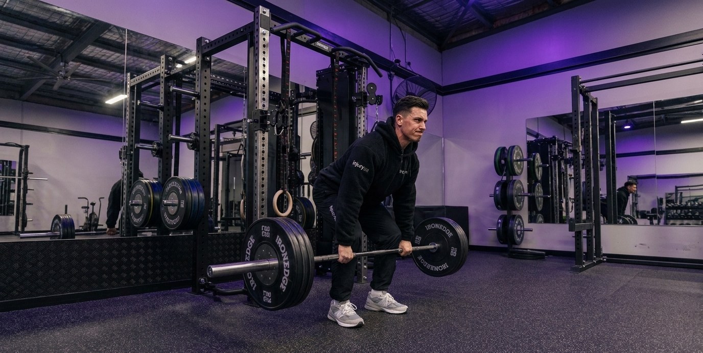
Then you sit with it for a week and start moving less, lifting nothing, and mentally writing off the gym. That reaction is completely understandable. It is also, for most people, the wrong response to what the scan actually shows.
Imaging is a picture of structure. Pain is an output of a nervous system responding to load, tissue sensitivity, sleep, stress, strength and history. The two overlap far less than most people assume, and the research on this is not new or controversial.
What the research actually found in people without pain
The clearest data comes from Brinjikji and colleagues (2015), who pooled 33 studies covering more than 3,000 people who had no back pain at all and put them through spinal imaging. The findings were striking.
- Disc degeneration was present in 37% of pain-free 20-year-olds and 96% of pain-free 80-year-olds.
- Disc bulges were present in 30% of pain-free 20-year-olds and 84% of pain-free 80-year-olds.
- Disc protrusions were present in 29% of pain-free 20-year-olds and 43% of pain-free 80-year-olds.
Read that again. Most pain-free adults in their fifties and sixties are walking around with disc bulges and degeneration they will never know about unless someone scans them.
This is not a recent discovery either. Boden and colleagues (1990) scanned asymptomatic volunteers and found that roughly a third had a substantial abnormality on MRI, with the rate rising sharply in those over 60. Nakashima and colleagues (2015) did the same thing in the neck with 1,211 healthy volunteers and found disc bulging in the large majority of them, across every age bracket.
In other words, degenerative changes on a spinal scan are closer to grey hair than to a fracture. They are a normal, near-universal feature of a lived-in body. They correlate with age far more reliably than they correlate with pain.
Why an early scan can sometimes make things worse
This is the part that surprises people. Getting imaged early for ordinary low back pain, in the absence of concerning features, is associated with worse outcomes, not better ones.
Webster and Cifuentes (2010) followed workers with acute low back pain and no red flag signs. Those who received early MRI had longer disability duration, higher costs and higher rates of surgery than those who did not, even after adjusting for severity. Chou and colleagues (2009), in a Lancet meta-analysis, found that routine imaging for low back pain did not improve pain, function or quality of life compared with usual care without imaging.
Why would a picture make things worse? Because a scary report changes behaviour. People who believe their spine is crumbling move less, guard more, avoid load and stop trusting their body. That deconditioning and fear-avoidance loop is one of the most reliable predictors of pain becoming persistent. The scan did not damage the back. The interpretation of the scan changed what the person did with their back.
When imaging genuinely matters
None of this means scans are useless. They are an important tool used at the right moment for the right reason. Imaging is clinically appropriate when there are red flags or when the result will actually change the plan, including:
- Suspected fracture, particularly after significant trauma or in the context of osteoporosis
- Signs suggestive of infection, inflammatory disease or malignancy, such as unexplained weight loss, fever, night pain or a relevant medical history
- Progressive neurological deficit, including worsening weakness, or any suspicion of cauda equina syndrome (saddle numbness, bladder or bowel changes), which is a medical emergency
- Severe, unremitting radicular pain that has not responded to a properly delivered course of conservative care and where surgical or injection options are being genuinely considered
A good assessment screens for all of this. If your history and examination point that way, imaging gets organised and the appropriate referral is made. What we avoid is scanning reflexively and then treating the report instead of the person in front of us.
Pain does not equal damage, and motion is medicine
Modern pain science has moved a long way from the plumbing model of the body. Pain is protective. It is the nervous system's best guess about threat, built from tissue input, previous experience, beliefs, sleep quality, stress load and how much capacity you currently have.
That is why two people with near identical scans can have completely different experiences. One has a disc bulge and plays Saturday footy. The other has the same disc bulge and cannot sit through a work meeting. The structural picture is the same. The load tolerance, the confidence and the context are not.
It is also why symptoms can improve substantially without a single structural change on imaging. The disc bulge is often still there when the pain has gone. What has changed is the sensitivity of the system and the capacity of the person to handle their daily loads.
This is where our approach starts. We find the cause. We fix the problem. We keep it away. The cause is rarely a word on a radiology report. It is usually a mismatch between what your body is being asked to do and what it is currently conditioned to do, layered on top of movement habits and beliefs that have narrowed over time.
Strength is the treatment, not the thing you do afterwards
If pain is largely a capacity problem, then building capacity is treatment. Not a bonus once you feel better. The treatment itself.
Owen and colleagues (2020) ran a network meta-analysis in the British Journal of Sports Medicine comparing exercise types for chronic low back pain and found resistance-based, motor control and Pilates style programs among the most effective for pain and physical function. Searle and colleagues (2015) reached a similar conclusion, with strength and coordination based programs producing meaningful reductions in chronic low back pain. Steffens and colleagues (2016) went further upstream and found that exercise, with or without education, reduced the risk of a new low back pain episode.
The pattern across the literature is consistent. Loading a back progressively and confidently tends to help. Protecting it indefinitely tends not to.
How we handle a scary scan report
When someone arrives at IAC with an MRI report and a lot of worry, the first job is a proper assessment: history, red flag screening, movement testing, strength and load tolerance, and a genuine conversation about what the findings do and do not mean for them. The scan is one piece of information sitting alongside everything else, not the verdict.
From there, you get one plan from one integrated team. We do not hand you between separate services with separate agendas. Hands-on treatment is used to reduce sensitivity and restore movement so that you can load properly, and the loading work is what builds the durability that keeps the problem away. At our Ravenhall clinic the treatment rooms and the rehab gym sit in the same building, which means the manual work and the strength work happen as part of the same session and the same conversation, not in two different places with two different stories.
Practically, that usually looks like graded exposure back into the movements you have been avoiding, progressive strength work for the hips, trunk and legs, and clear education on why hurt does not automatically mean harm. Most people with ordinary mechanical low back pain and even many with genuine sciatica improve with well-delivered conservative care over 6 to 12 weeks, though timelines vary considerably between individuals and some will need imaging or specialist input along the way.
What to take from your report
If your scan mentions degeneration or a bulging disc, ask three questions. Does this finding match my symptoms and my examination? Would treating this finding change what I do next? What is my body currently unable to tolerate, and how do we build that back?
Those questions move the focus from a static image to something you can actually influence. The disc bulge is not going to be argued out of existence. Your strength, your movement confidence and your tolerance for load absolutely can change.
If you have been sitting on a scan report and moving less because of what it says, it is worth having it properly put in context and turning it into a plan. Explore our treatment services, or book a consultation at any of our Melbourne clinics in Ravenhall or Altona to discuss your situation.
Related conditions & services
Ready to move better?
Book an appointment with our team and let's get to the root cause of what's bothering you.